If you’ve ever noticed a small lump on your scalp or skin, you’ve likely wondered if it’s harmless or needs medical attention.
Cysts are among the most common non-cancerous skin lumps seen in dermatology.
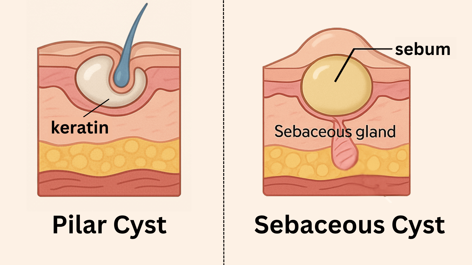
Two types often mistaken for each other are the pilar cyst and the sebaceous cyst, also known as an epidermoid cyst.
Though both are generally harmless, their origins, locations, and treatments differ.
Recognizing the difference between pilar and sebaceous cysts helps determine when medical care or removal may be necessary.
This blog explains how each cyst forms, what they look like, and the safest ways to manage or remove them.
What is a Pilar Cyst?
A pilar cyst (trichilemmal cyst) forms from the outer root sheath of a hair follicle.
It’s most commonly found on the scalp and often runs in families, suggesting a genetic component.
Common features of a pilar cyst:
- Round, firm, and smooth lump beneath the scalp.
- Usually painless and slow-growing.
- No visible opening (punctum).
- Filled with keratin, a white, firm protein substance.
A pilar cyst may appear singly or in clusters and can occasionally become irritated or inflamed.
While generally benign, some people choose pilar cyst removal to prevent discomfort or for cosmetic reasons.
What is a Sebaceous (Epidermoid) Cyst?
A sebaceous cyst, often called an epidermoid cyst, develops when a sebaceous gland or hair follicle becomes blocked, trapping oil and dead skin cells beneath the surface.
Common characteristics of a sebaceous cyst:
- Softer than a pilar cyst.
- Has a small central punctum (pore).
- It may produce a yellowish, oily discharge if ruptured.
- Commonly found on the face, neck, chest, or back.
Sebaceous cyst treatment may not always be necessary unless the cyst becomes red, swollen, or infected.
Difference Between Pilar and Sebaceous Cysts
Both pilar cysts and sebaceous cysts are benign, but they differ in their origin, texture, and content.
| Feature | Pilar Cyst | Sebaceous (Epidermoid) Cyst |
|---|---|---|
| Origin | Outer root sheath of a hair follicle | Blocked sebaceous gland or follicle |
| Content | Keratin (white, thick material) | Sebum (oily yellowish fluid) |
| Texture | Firm and smooth | Softer, may fluctuate |
| Location | Scalp and hair-bearing areas | Face, neck, chest, or back |
| Punctum | Absent | Usually visible |
| Genetic Link | Often hereditary | Not hereditary |
| Infection Risk | Rare | More likely |
| Treatment | Surgical excision | Excision or drainage if inflamed |
These differences help identify if a lump is a scalp cyst (often a pilar cyst) or a sebaceous cyst elsewhere on the body.
Are Pilar and Sebaceous Cysts Dangerous?
Both pilar cysts and sebaceous (epidermoid) cysts are benign, meaning they are not cancerous.
They develop slowly and rarely lead to serious health problems.
However, complications can arise if the cyst becomes infected, ruptures under the skin, or causes discomfort due to size or location.
Malignant change (becoming cancerous) is extremely rare, but it’s best to have any cyst that changes rapidly in size, color, or texture checked by a professional.
With proper medical assessment and timely cyst removal, recovery is straightforward, and recurrence is uncommon.
|
Toxicity to Pets Disclaimer Neither pilar cysts nor sebaceous cysts in humans are contagious or harmful to pets. However, pets can develop their own sebaceous cysts that may require veterinary treatment. Keep human antiseptics or topical medications away from animals, as some ingredients can be toxic if licked or ingested. |
Causes for Pilar and Sebaceous Cysts
Knowing the causes behind pilar cysts and sebaceous cysts helps identify what triggers them and how to manage or prevent new ones from forming.
Each has distinct origins depending on where and how it develops.
Pilar Cyst Causes
Pilar cysts develop when excess keratin, a natural protein found in hair, accumulates within the hair follicle.
They often have a genetic link, running in families where multiple members experience similar cysts.
Minor scalp injuries or repeated irritation can also contribute to their formation.
Sebaceous Cyst Causes
Sebaceous cysts form when the sebaceous glands become blocked, typically due to oil buildup, injury, or skin trauma such as acne.
Hormonal fluctuations or naturally oily skin can increase the likelihood of gland obstruction.
In some cases, frequent sun exposure can stimulate sebaceous gland activity, leading to a higher risk of cyst formation.
Both types remain non-cancerous but can occasionally become infected, requiring prompt medical attention.
Symptoms and Appearance of Pilar and Sebaceous Cysts
Both pilar cysts and sebaceous cysts appear as lumps under the skin, but they differ slightly in firmness, texture, and placement.
Knowing what to look for helps identify which type you might have.
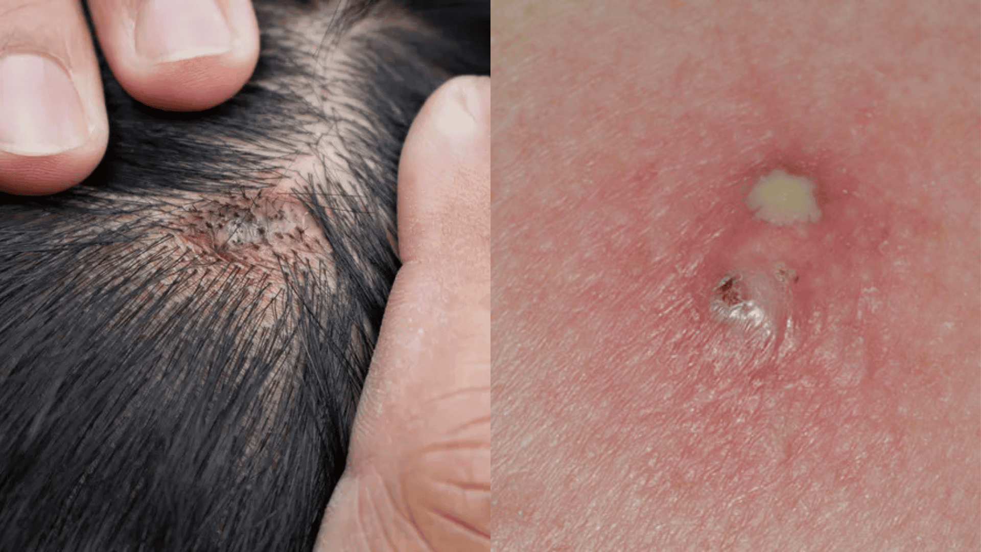
Pilar cyst:
- Feels firm and rubbery.
- No visible pore.
- Commonly seen on the scalp or behind the ears.
Sebaceous cyst:
- Softer and may have a small pore at the center.
- May leak a cheesy or oily discharge when ruptured.
- Found on the face, upper back, or chest.
In both cases, redness, tenderness, and swelling may indicate infection.
Medical treatment or cyst removal prevents further inflammation.
When to See a Doctor for a Pilar or Sebaceous Cyst?
Most pilar and sebaceous cysts are harmless, but medical attention may be needed if a lump grows quickly, becomes painful, or shows signs of infection such as redness, swelling, or discharge.
If the cyst causes discomfort or changes appearance, it’s best not to ignore it.
Persistent or recurring cysts, especially those difficult to identify, also warrant a checkup.
A dermatologist can diagnose the type of cyst through examination or imaging and recommend safe removal options suited to your condition.
Diagnosis of Pilar and Sebaceous Cysts
Diagnosing a pilar cyst or sebaceous cyst is usually straightforward, but doctors may use imaging or lab tests to confirm the type, rule out infection, and ensure the lump isn’t something more serious.
A correct diagnosis helps guide safe and effective treatment.
1. Clinical examination

A dermatologist begins by carefully inspecting and feeling the lump.
The cyst’s location, texture, and appearance often reveal its type.
Firm and smooth lumps on the scalp suggest a pilar cyst, while softer bumps with a visible pore often indicate a sebaceous cyst.
2. Dermatoscopy
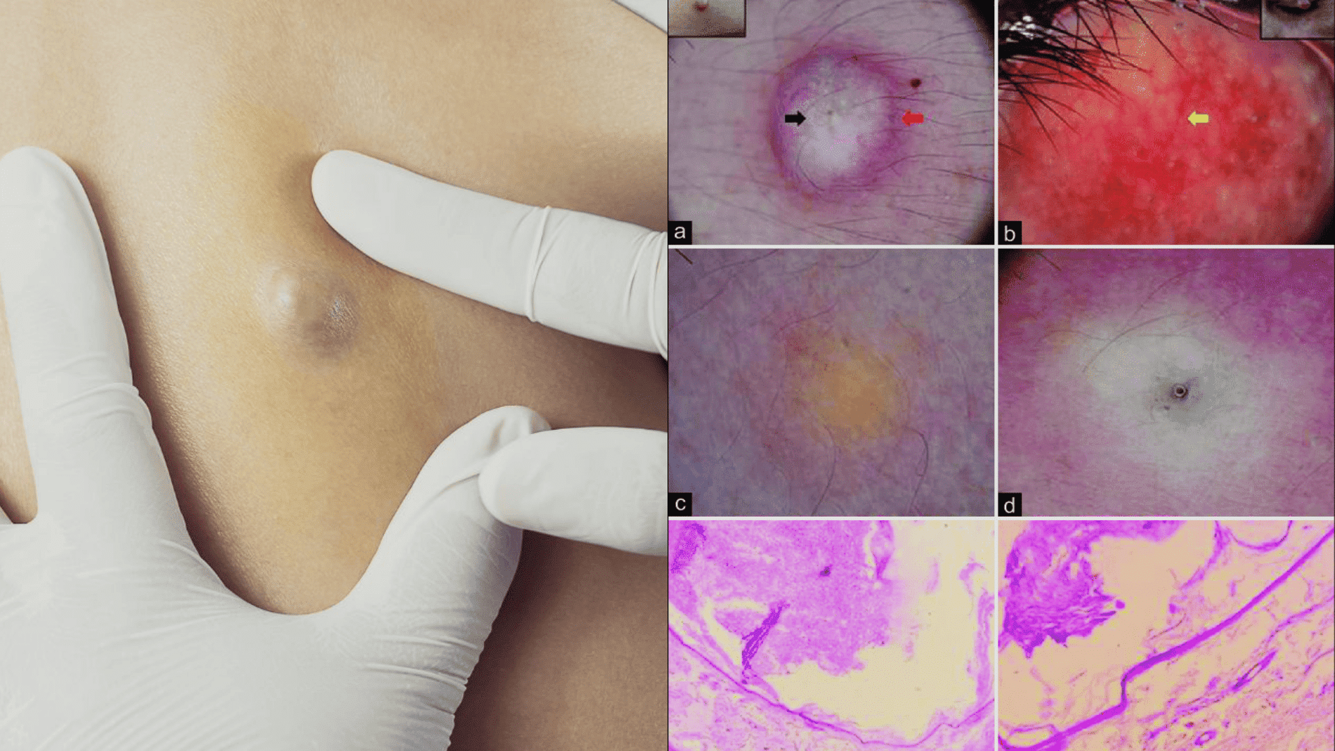
A handheld magnifier called a dermatoscope allows the doctor to view surface details such as the presence of a punctum or color variation.
This tool helps distinguish cysts from lipomas, warts, or other skin growths.
3. Ultrasound

An ultrasound scan gives a clear picture of the cyst’s internal structure, confirming if it contains keratin or sebum.
It also helps assess cyst depth, nearby blood vessels, and inflammation useful before cyst removal.
4. Biopsy

If the cyst appears atypical, a small tissue sample may be taken for histopathological testing.
This confirms if the cyst is pilar or sebaceous (epidermoid) and rules out rare skin tumors or infections.
5. Infection check
If redness or pain is present, the doctor might collect a swab from any discharge to identify bacteria.
This ensures that proper antibiotics are prescribed before removal.
Both pilar cysts and sebaceous cysts are benign. Malignant changes are extremely rare, but early examination is still advised for any cyst that grows quickly, becomes painful, or changes in color or texture.
Treatment Options for Pilar and Sebaceous Cysts
Treatment depends on the cyst’s size, location, and whether it’s causing symptoms.
While small cysts can sometimes be monitored, most pilar cysts and sebaceous cysts respond best to simple removal procedures.
1. Observation: If the cyst is small, painless, and not infected, no immediate treatment is necessary. Many patients simply monitor the lump over time.
2. Surgical Removal: The most reliable pilar cyst treatment and sebaceous cyst removal method is surgical excision under local anesthesia.
- The entire cyst wall and contents are removed through a small incision.
- Procedure time: 15–30 minutes.
- Recurrence is rare if fully excised.
3. Drainage and Medication: If infection occurs:
- Incision and drainage may provide relief.
- Antibiotics treat bacterial infections.
- Steroid injections may help reduce swelling before removal.
Proper aftercare ensures quick healing and prevents recurrence.
Aftercare Following Cyst Removal
Post-procedure care plays an important role in how well the skin heals after pilar cyst removal or sebaceous cyst removal.
Here’s how to care for the treated area:
- Keep the area clean and dry: Gently cleanse the site with mild soap and water once daily. Pat dry, avoid rubbing.
- Avoid pressure and irritation: Do not squeeze, scratch, or pick at the healing wound, as this can delay recovery or introduce bacteria.
- Apply medication as directed: Use any prescribed antiseptic creams or antibiotic ointments to protect the area.
- Protect the site from moisture: Avoid swimming or long showers until stitches are removed.
- Stitch care: If sutures were placed, they are usually removed within 7–10 days. Dissolvable stitches will naturally break down within a few weeks.
Expected recovery: Most patients heal within one to two weeks, with minimal scarring. For facial cysts, applying a silicone-based scar gel or sheet can help smooth and fade marks.
How to Prevent Cysts?
While not all cysts can be prevented, especially if they’re genetic, certain lifestyle habits can lower the risk of pilar cysts or sebaceous cysts forming or returning.
- Wash regularly to remove oil buildup and dead skin cells that can block follicles.
- Avoid picking or squeezing lumps.
- Treat acne promptly, as managing acne and oily skin prevents clogged pores that can lead to sebaceous cysts.
- Choose gentle products; use mild, non-comedogenic skincare and hair products to minimize irritation.
- If you’ve had a pilar cyst before, regular checks can catch small cysts before they enlarge or get infected.
If cysts run in your family, early consultation with a dermatologist can help plan ongoing pilar cyst treatment and reduce recurrence.
Conclusion
Knowing the difference between pilar and sebaceous cysts helps in recognizing which type you might have and the best treatment approach.
A pilar cyst grows from a hair follicle, commonly on the scalp, while a sebaceous (epidermoid) cyst arises when an oil gland becomes blocked.
If you notice a persistent lump or cyst, consult a dermatologist for proper diagnosis, reassurance, and lasting relief.
Have you ever dealt with a pilar cyst or sebaceous cyst?
Share your experience or questions in the comments below!
Frequently Asked Questions
Can Pregnancy Cause Sebaceous Cysts?
Yes. Hormonal changes during pregnancy can increase oil production, which may lead to sebaceous cyst formation. These cysts are harmless and can be removed safely if they cause discomfort.
Can a Cyst Feel Rock Hard?
Yes. A pilar cyst or an older sebaceous cyst can feel quite hard due to keratin or sebum buildup. Though firm, it’s usually benign and may need cyst removal if painful or irritated.
Can a Sebaceous Cyst Last for Years?
Yes. Some sebaceous cysts can remain unchanged for several years. However, if the cyst enlarges, becomes red, or causes discomfort, sebaceous cyst removal is the best treatment option.