
Leg vein symptoms may appear simple at first glance, yet the source is often hidden below the skin. Swelling, heaviness, aching, and visible varicose veins may point to chronic venous insufficiency, but surface changes tell only part of the story. Ultrasound imaging gives vein doctors a live map of blood flow in the legs. That map helps them confirm the problem, measure its extent, and choose minimally invasive treatments with greater accuracy.
A Clearer Starting Point
Before any treatment plan is set, doctors need to see which leg veins are not moving blood well. At Metro Vein Centers, ultrasound imaging helps physicians examine vein function, valve failure, and blood flow patterns in real time. This added detail supports a more exact diagnosis for varicose veins, spider veins, and chronic venous insufficiency, while also helping to rule out serious concerns such as deep vein thrombosis.
What Ultrasound Shows
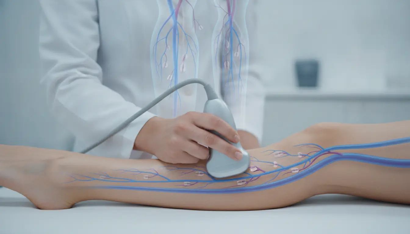
Ultrasound creates images of leg veins without surgery or radiation. It shows whether the venous valves are closing as they should. The scan can also reveal reflux, which means blood is moving in the wrong direction. That backward flow often causes pressure to build in the legs. Knowing where reflux begins helps doctors avoid guesswork during treatment planning.
Why Surface Exams Fall Short
A physical exam can reveal bulging veins or skin changes, but it cannot show full vein function. Some damaged veins sit deeper in the leg and remain hidden from view. Symptoms may feel severe even when surface veins appear mild. Ultrasound fills that gap by showing the vein network under the skin, along with the blood flow that drives symptoms.
Mapping the Exact Problem Areas
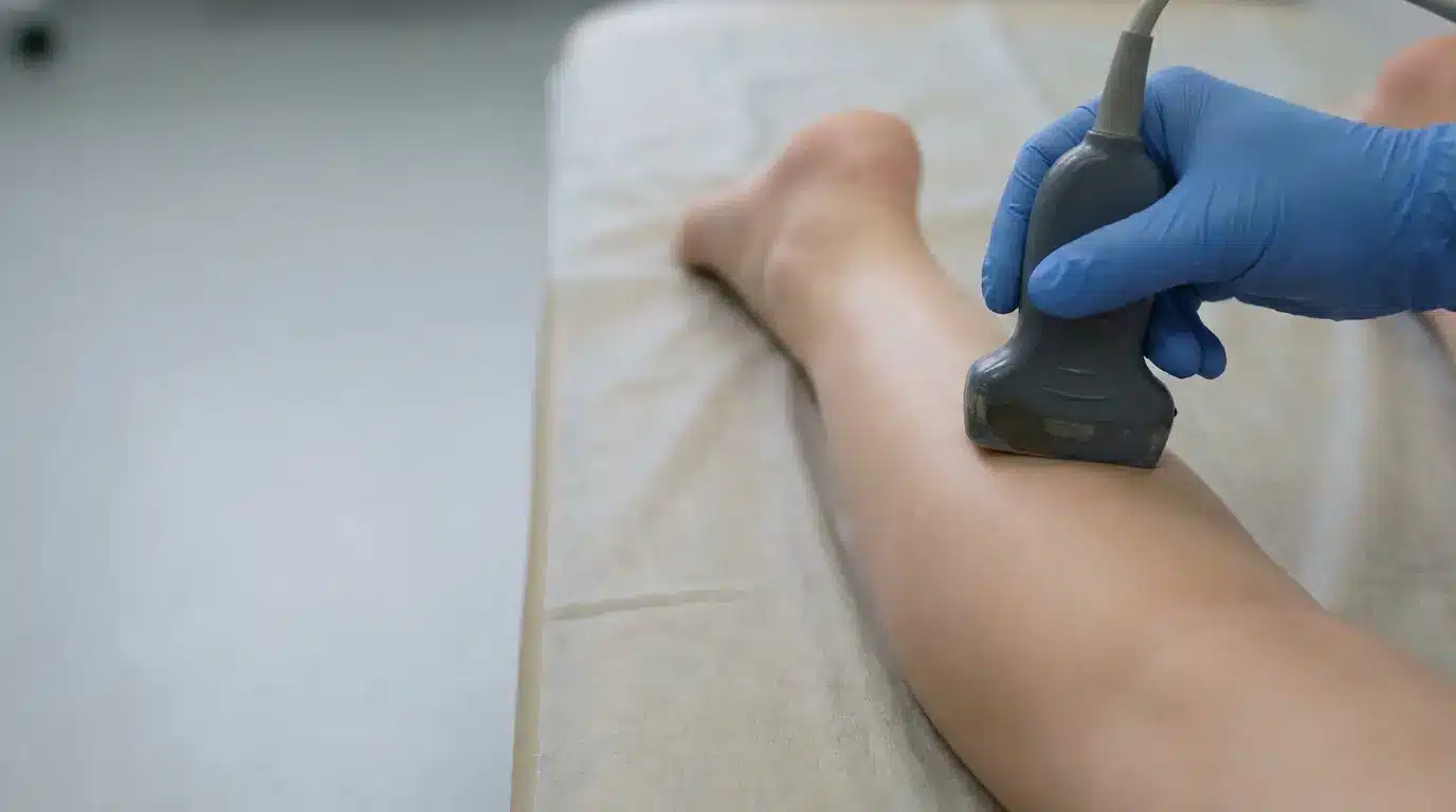
Precise care depends on knowing which veins are feeding the problem. Ultrasound helps doctors trace the path of faulty blood flow through the leg. That matters because visible veins are often a result, not the main source. Treating the wrong branch may leave symptoms in place. A detailed map supports a plan that targets the true cause.
Matching Findings to Treatments
Once doctors identify the source of reflux, they can choose the best minimally invasive treatments for that pattern. Some patients may benefit from sclerotherapy for smaller veins. Others may need radiofrequency ablation, adhesive closure, or another image-guided option for larger veins. Ultrasound helps match the treatment to the vein size, location, and blood flow issue.
Supporting Safer Decisions
Ultrasound also improves safety during planning. It helps doctors confirm where healthy veins should be preserved and where diseased veins can be treated. That distinction matters because leg circulation depends on a working network. Good imaging lowers the chance of treating an area that is not driving symptoms. Better planning often means fewer surprises on treatment day.
Guiding Treatment in Real Time
In many cases, ultrasound remains useful after planning is complete. During certain treatments, doctors use it to guide placement accurately within the leg vein. Real-time imaging helps them follow the target vein and monitor progress as treatment moves forward. That extra visibility supports a careful approach, which can improve comfort and help produce a more focused result.
Helping Patients Understand Their Condition
Seeing the scan can also help patients understand why symptoms are happening. A doctor can point out reflux, enlarged veins, or valve failure during the visit. That visual explanation often makes the treatment plan easier to follow. Patients may feel more prepared when they can connect leg pain or swelling to a clear image rather than relying on description alone.
Useful for Early and Advanced Cases
Ultrasound is valuable across many stages of vein disease. Some patients seek help when spider veins first appear. Others wait until heaviness, cramping, or swelling becomes hard to ignore. In both cases, imaging can show whether the issue is cosmetic, medical, or a mix of both. Early answers may help prevent ongoing pressure from causing broader leg discomfort.
Conclusion
Precise vein care starts with clear information, and ultrasound provides that foundation. By showing blood flow, valve function, and hidden reflux in the legs, it allows vein doctors to diagnose problems with more confidence and plan minimally invasive treatments that fit each patient’s needs. That level of detail matters for both comfort and results. For people dealing with varicose veins, spider veins, or chronic venous insufficiency, ultrasound helps turn symptoms into a focused treatment strategy.